Technology and Diagnostics
Research projects
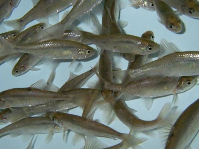
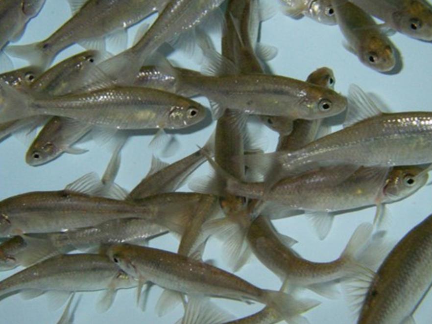
Better fish tagging
ODFW researchers are evaluating a range of fish marking methods using Oregon chub. Visible Implant Elastomer (ViE: an injectable colored plastic) was found to produce an effective mark. Vie was used successfully to document fish migration patterns in 3 river sub-basins. The VIE marks persisted for at least 3 years. Resource management and recovery operations need a way to track the movement of free-range fish. This requires a method of tagging or marking the fish-- however, reliable methods to track smaller fish are lacking.
Bangs, B.L., Falcy, M.R., Scheerer, P.D. and Clements, S. 2013. Comparison of three methods for marking a small floodplain minnow. Anim. Biotelemetry 1(18).
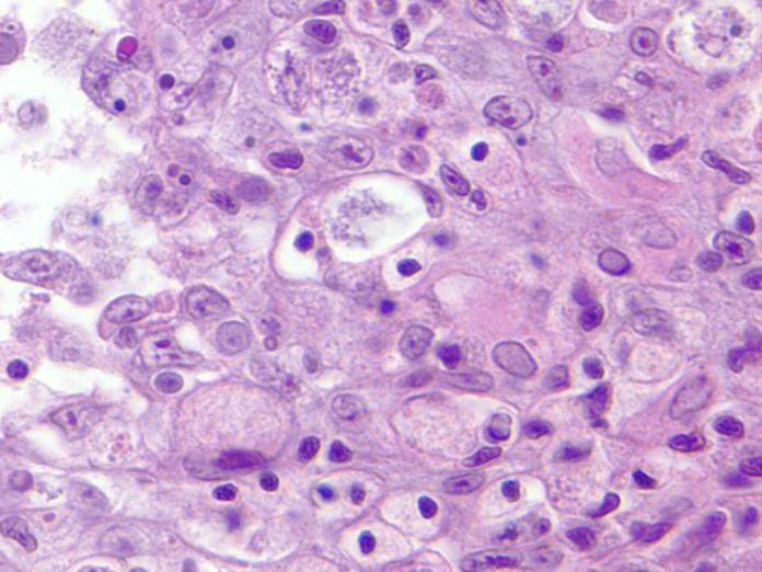

Detecting koi herpes virus
Dr. Ling Jin's lab (OSU Carlson College of Veterinary Medicine) has been studying Koi herpes virus for several years at the AAHL. Since the virus can lie dormant ornamental koi owners may not know their fish are infected. KHV is a virus that infects ornamental koi and common carp worldwide. Like most viral infections there is currently no treatment for KHV. Infected fish can show gill lesions, sunken eyes or a notched nose. Transport to international koi shows and displays in communal aquaria have spread the virus worldwide. In an effort to identify a marker that will lead to a quick and easy test for owners, Dr. Jin and Dr. Reed demonstrated that KHV can hide in the fish's white blood cells.
Reed, A., Lin, L., Ostertag-Hill, C., Wang, Q., Wu, Z., Miller-Morgan, T., and Jin, L. 2017. Detection of ORF6 protein associated with latent KHV infection. Virology 500:89-90.
Prescott, M., Reed, A., Jin, L., and Pastey, M. 2016. Rapid detection of Cyprinid herpesvirus 3 in latently infected koi by recombinase polymerase amplification. J. of Aquatic Animal Health 28:173-180.
Microbial source tracking
Lauren Brooks (Kate Field lab; OSU Microbiology) studied the rate of degradation of agricultural fecal input in a water body and the microbial communities responsible for that breakdown. Microbial Source Tracing (MST) is an attempt to determine non-point sources of fecal contamination. A major limitation to implementation of this field is the poor understanding of persistence of pathogens and indicator bacteria in the environment. The study tested the rate at which agricultural fecal contamination is broken down by microbes in water bodies like rivers and ponds. The research originally stemmed from a concern that there was not always an effective way to differentiate between the sources of fecal contamination. The data has been used to develop a ratio model which will be useful for allocation of pollutant sources leading to targeted pollution mitigation efforts.
Brooks, L.E. and Field, K.G. 2016. Bayesian meta-analysis to synthesize decay rate constant estimates for common fecal indicator bacteria. Water Res. 104.
Brooks, L.E. and Field, K.G. 2017. Global model fitting to compare survival curves for faecal indicator bacteria and ruminant-associated genetic markers. J. Appl. Microbiol. ????
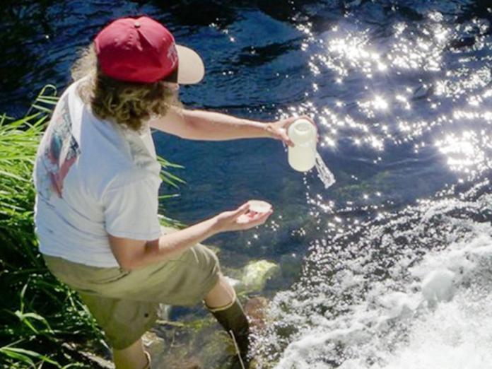

Pathogen detection in water
Dr. Jerri Bartholomew (OSU Microbiology) used quantitative PCR to detect pathogens in aquatic ecosystems. Water sampling is a more recent approach to test for aquatic pathogens such as bacteria and myxozoan parasites. Water is collected by hand or with an automatic sampler. Samples are filtered and the DNA is extracted. Molecular methods such as quantitative PCR are used to determine the identity and abundance of pathogens. Data is usally obtained more quickly than with sentinel fish exposure data, and has a higher detection threshold. A combined approach of water sampling and sentinel fish exposures is used to relate abundance of waterborne stages with fish health. Using PCR, the genotype of the parasite present in the water can also be identified which helps determine the risks to specific fish species.
Hallett, S.L. and Bartholomew, J.L. 2006. Application of a real-time PCR assay to detect and quantify the myxozoan parasite Ceratomyxa shasta in river water samples. Dis. Aquat. Organ. 71(2):109-118.
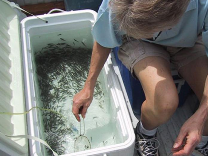
Sentinel fish for pathogen detection

Sentinel fish for pathogen detection
Dr. Jerri Bartholomew (OSU Microbiology) in collaboration with the Bureau of Reclamation are using "sentinel" fish to detect pathogens in aquatic ecosystems. Cages of live, pathogen free fish, are placed in particular locations at different times of the year. After 3-7 days of exposure, the fish are transported to the AAHL where they are held in clean water at selected temperatures and monitored daily for signs of disease. Sentinel fish can alert us to where and when certain pathogens are present, and which fish species/stocks are susceptible to different pathogens.
View full study
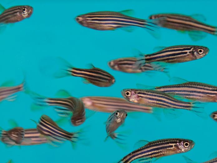

Zebrafish in outer space
Dr. Jack Higginbotham (Oregon Space Grant consortium) and Dr. Jan Spitsbergen (Microbiology) are collaborating on a NASA funded project to develop a zebrafish model for radiation-induced cataracts. Pilots, astronauts, and those exposed to high levels of ultraviolet light or ionizing radiation are at high risk of developing cataracts at a young age. Zebrafish can be ?????????????????? galactic cosmic rays when on extended space missions.
In this study, zebrafish have been exposed to gamma irradiation at Brookhaven National Lab on Long Island, New York, as well as simulated high energy galactic cosmic ray exposure. In collaboration with Dr. Kirsten Lampa (OHSU) and Dr. Sarah Maxwell (Opthalmic Veterinarian) proteomic studies (proteins) of the lens of the eyes should provide data on threshold limits of galactic cosmic rays on zebrafish and hence to astronauts.
Higginbotham, J., Spitsbergen, J., Maxwell, S., Jardine, J., Guida, P., Hudecek, C., Parker, H., Folk, L., Milston-Clements, R. and Muller, G. 2019. Investigation of link between zebrafish cataract formation from exposure to galactic cosmic radiation and 137 Cs gamma rays.